
To determine whether you may have a deep vein thrombosis (DVT) or pulmonary embolism (PE), your doctor will ask you questions about your current symptoms and obtain your medical history to better assess your risk factors.
A physical examination will be done to check for extremity swelling, tenderness and skin discoloration. If your symptoms, medical history and physical examination suggest that a blood clot is likely, testing will be done which may include:

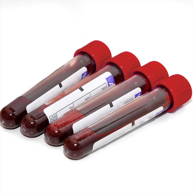
A blood test can be used to rule out presence of a DVT. If the D-dimer test is negative and you are determined to have a low-risk for DVT (based upon the history and physical examination), further testing with an imaging study to rule out a blood clot may not be needed. However, if the suspicion that you have a blood clot is intermediate or high, an imaging study needs to be done.

A Doppler ultrasound is a painless and noninvasive test used to diagnose DVT. During a Doppler ultrasound, sound waves are used to generate pictures of the blood vessels. In most cases, Doppler ultrasound is the preferred test to diagnose DVT.
A contrast venogram is often reserved for situations in which a Doppler ultrasound is not feasible. During contrast venogram, a catheter is inserted into a vein and dye is injected, allowing your doctor to see the vein with an x-ray.
A magnetic resonance imaging (MRI) uses a strong magnet to create an image of inside the body. MRI is reserved for situations in which a contrast venogram cannot be performed.
Computer tomography (CT) venography or MRI venography are the preferred tests to look at blood clots in the pelvis or the abdomen.